Welcome to the Amira-Avizo Software Use Case Gallery
Below you will find a collection of use cases of our 3D data visualization and analysis software. These use cases include scientific publications, articles, papers, posters, presentations or even videos that show how Amira-Avizo Software is used to address various scientific and industrial research topics.
Use the Domain selector to filter by main application area, and use the Search box to enter keywords related to specific topics you are interested in.
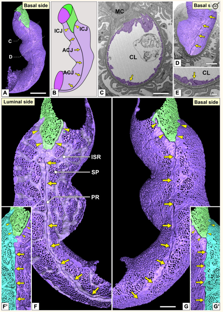
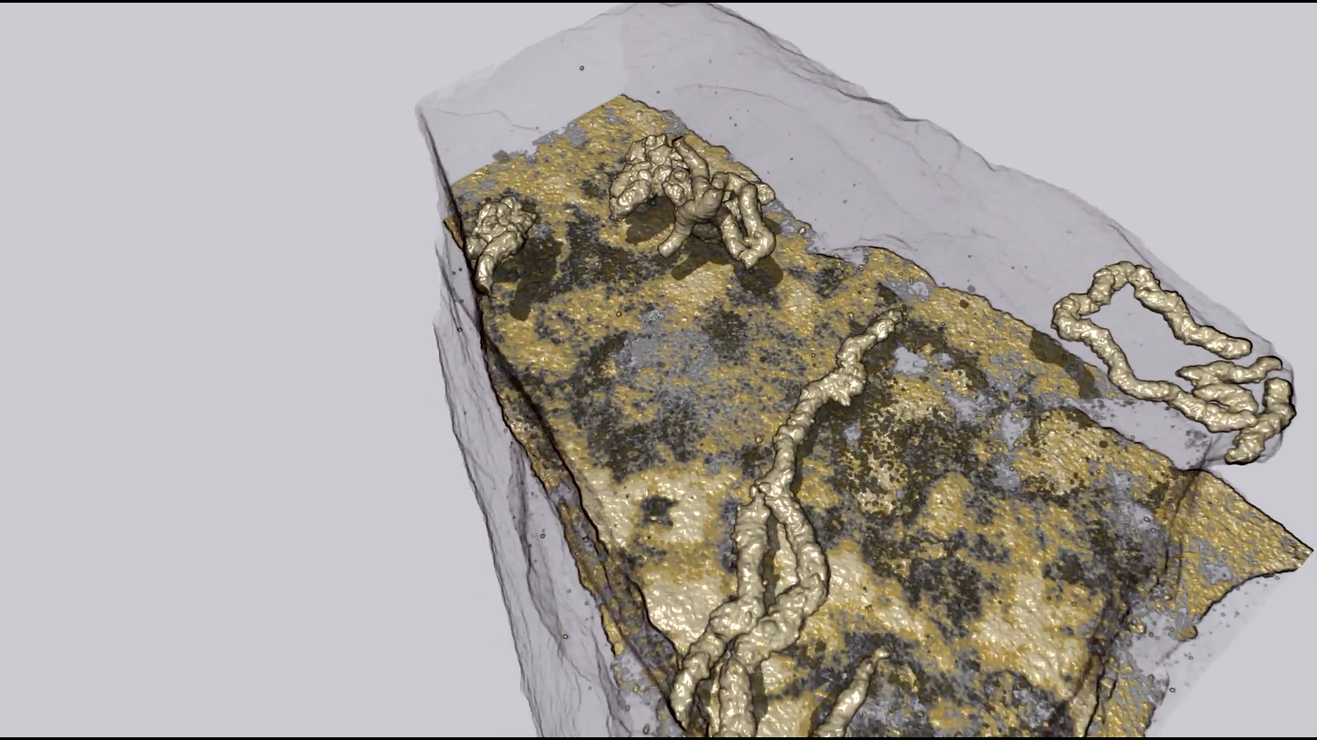
Three-Dimensional Architecture of Glomerular Endothelial Cells Revealed by FIB-SEM Tomography
Focused-ion beam-scanning electron microscopic (FIB-SEM) tomography enables easier acquisition of a series of ultrastructural, sectional images directly from resin-embedded biological samples. In this study, to clarify the three-dimensional (3D) architecture of glomerular endothelial cells (GEnCs) in adult rats, we manually extracted GEnCs from serial FIB-SEM images and reconstructed them on an Amira reconstruction software. The luminal and basal surface structures were clearly visualized in ... Read more
Yuto Kawasaki, Yasue Hosoyamada, Takayuki Miyaki, Junji Yamaguchi, Soichiro Kakuta, Tatsuo Sakai, and Koichiro Ichimura
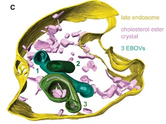
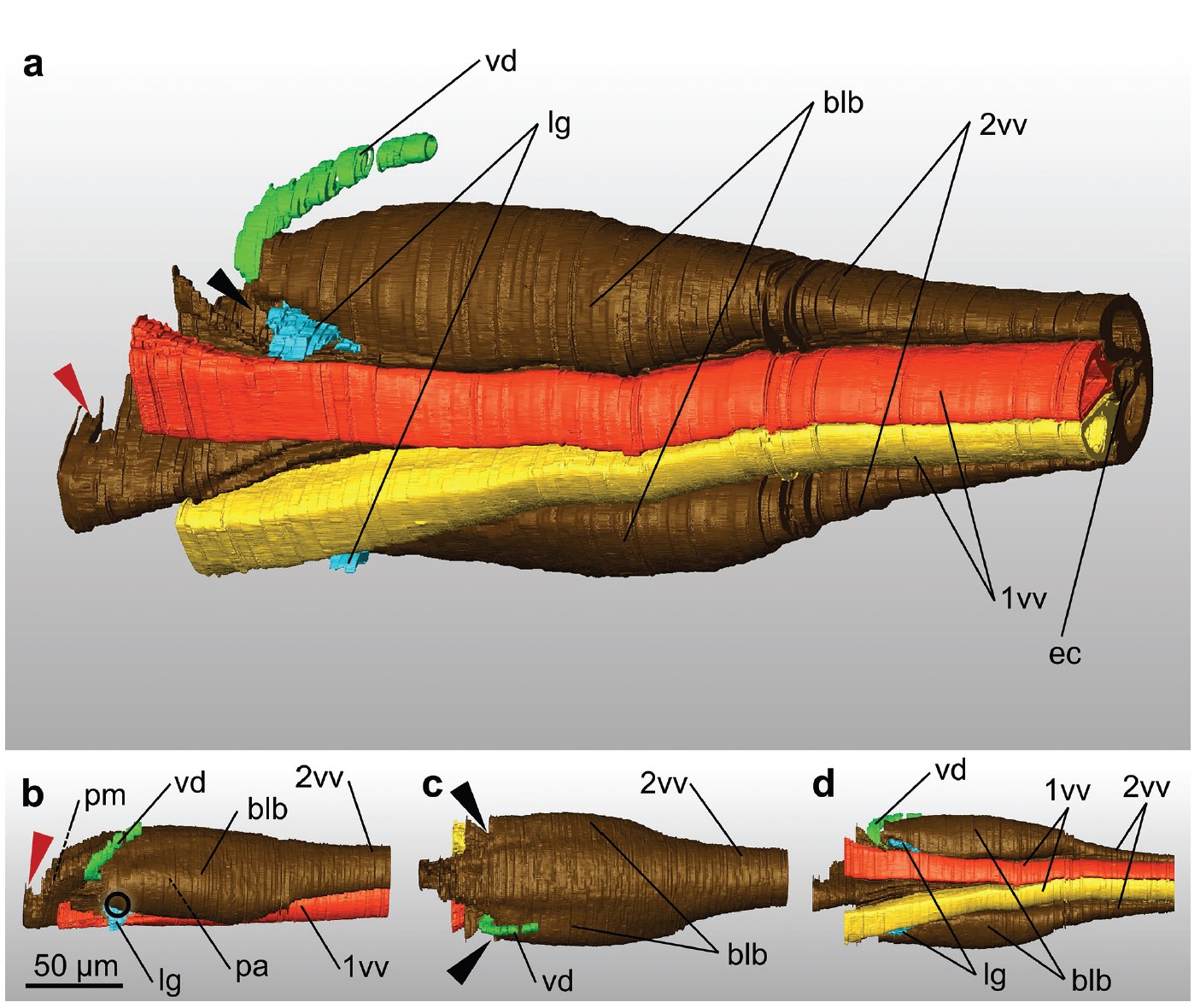
The Ebola virus VP40 matrix layer undergoes endosomal disassembly essential for membrane fusion
Ebola viruses (EBOVs) assemble into filamentous virions, whose shape and stability are determined by the matrix viral protein 40 (VP40). Virus entry into host cells occurs via membrane fusion in late endosomes; however, the mechanism of how the remarkably long virions undergo uncoating, including virion disassembly and nucleocapsid release into the cytosol, remains unknown. Here, we investigate the structural architecture of EBOVs entering host cells and discover that the VP40 matrix disassem... Read more
Sophie L Winter, Gonen Golani, Fabio Lolicato, Melina Vallbracht, Keerthihan Thiyagarajah, Samy Sid Ahmed, Christian Lüchtenborg, Oliver T Fackler, Britta Brügger, Thomas Hoenen, Walter Nickel Ulrich S Schwarz, Petr Chlanda
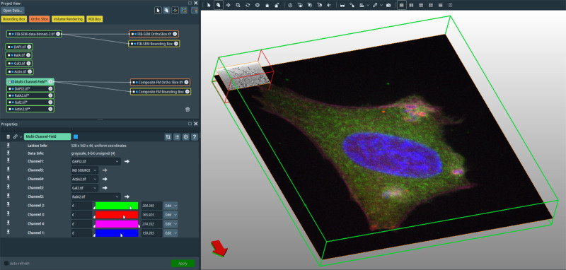
In recent years new methodologies and workflow pipelines for acquiring correlated fluorescence microscopy and volume electron microscopy datasets have been extensively described and made accessible to users of different levels. Post-acquisition image processing, and particularly correlation of the optical and electron data in a single integrated three-dimensional framework can be key for extracting valuable information, especially when imaging large sample volumes such as whole cells or tissu... Read more
Allon Weiner
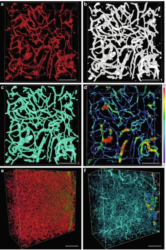
Despite advances in imaging, image-based vascular systems biology has remained challenging because blood vessel data are often available only from a single modality or at a given spatial scale, and cross-modality data are difficult to integrate.
Therefore, there is an exigent need for a multimodality pipeline that enables ex vivo vascular imaging with magnetic resonance imaging, computed tomography and optical microscopy of the same sample, while permitting imaging with complementary c... Read more
Akanksha Bhargava, Benjamin Monteagudo, Priyanka Kushwaha, Janaka Senarathna, Yunke Ren, Ryan C. Riddle , Manisha Aggarwal and Arvind P. Pathak
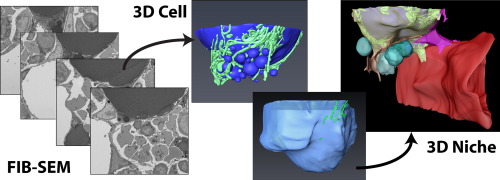
Characterization of the bone marrow adipocyte niche with three-dimensional electron microscopy
Unlike white and brown adipose tissues, the bone marrow adipocyte (BMA) exists in a microenvironment containing unique populations of hematopoietic and skeletal cells.
To study this microenvironment at the subcellular level, we performed a three-dimensional analysis of the ultrastructure of the BMA niche with focused ion beam scanning electron microscopy (FIB-SEM). This revealed that BMAs display hallmarks of metabolically active cells including polarized lipid deposits, a dense mitoch... Read more
Hero Robles, SungJae Park, Matthew S. Joens, James A.J. Fitzpatrick, Clarissa S. Craft, Erica L. Scheller
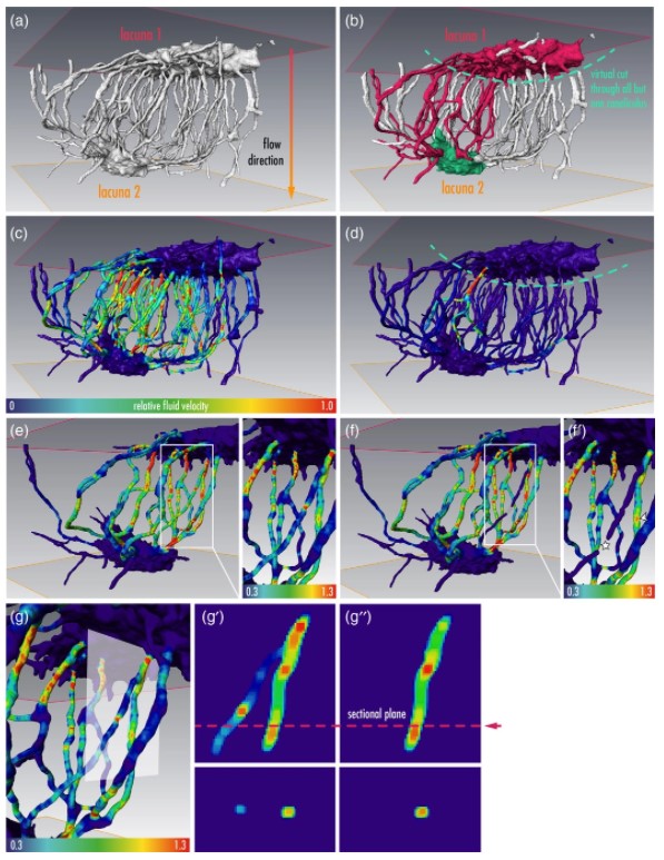
Cortical bone is permeated by a system of pores, occupied by the blood supply and osteocytes. With ageing, bone mass reduction and disruption of the microstructure are associated with reduced vascular supply. Insight into the regulation of the blood supply to the bone could enhance the understanding of bone strength determinants and fracture healing. Using synchrotron radiation-based computed tomography, the distribution of vascular canals and osteocyte lacunae was assessed in murine cortica... Read more
J.A. Núñez; A. Goring; B. Javaheri; H. Razi; D. Gomez-Nicola; E. Hesse; A.A. Pitsillides; P.J. Thurner; P. Schneider; E. Clarkin
Cryo-STEM mapping of solid–liquid interfaces and dendrites in lithium-metal batteries
Solid–liquid interfaces are important in a range of chemical, physical and biological processes but are often not fully understood owing to the lack of high-resolution characterization methods that are compatible with both solid and liquid components. For example, the related processes of dendritic deposition of lithium metal and the formation of solid–electrolyte interphase layers are known to be key determinants of battery safety and performance in high-energy-density lithium-metal bat... Read more
Michael J. Zachman, Zhengyuan Tu, Snehashis Choudhury, Lynden A. Archer & Lena F. Kourkoutis
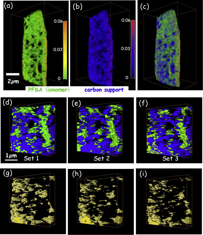
4D imaging – the three-dimensional distributions of chemical species determined using multi-energy X-ray tomography – of cathode catalyst layers of polymer electrolyte membrane fuel cells (PEM-FC) has been measured by scanning transmission x-ray microscopy (STXM) spectro-tomography at the C 1s and F 1s edges. In order to monitor the effects of radiation damage on the composition and 3D structure of the perfluorosulfonic acid (PFSA) ionomer, the same volume was measu... Read more
Juan Wu, Lis G.A.Melo, Xiaohui Zhu, Marcia M.West, Viatcheslav Berejnov, Darija Susac, Juergen Stumper, Adam P.Hitchcock
Paleozoic Nymphal Wing Pads Support Dual Model of Insect Wing Origins
The appearance of wings in insects, early in their evolution [1], has been one of the more critical innovations contributing to their extraordinary diversity. Despite the conspicuousness and importance of wings, the origin of these structures has been difficult to resolve and represented one of the “abominable mysteries” in evolutionary biology [2]. More than a century of debate has boiled the matter down to two competing alternatives—one of wings representing an extension of the thorac... Read more
Department of Zoology, Faculty of Science, Charles University, Praha, Czech Republic and al.
Organism motility in an oxygenated shallow-marine environment 2.1 billion years ago
Evidence for macroscopic life in the Paleoproterozoic Era comes from 1.8 billion-year-old (Ga) compression fossils [Han TM, Runnegar B (1992) Science 257:232–235; Knoll et al. (2006) Philos Trans R Soc Lond B 361:1023–1038], Stirling biota [Bengtson S et al. (2007) Paleobiology 33:351–381], and large colonial organisms exhibiting signs of coordinated growth from the 2.1-Ga Francevillian series, Gabon. Here we report on pyritized string-shaped structures from... Read more
Abderrazak El Albani, M. Gabriela Mangano, Luis A. Buatois, Stefan Bengtson, Armelle Riboulleau, Andrey Bekker, Kurt Konhauser, Timothy Lyons, Claire Rollion-Bard, Olabode Bankole, Stellina Gwenaelle Lekele Baghekema, Alain Meunier, Alain Trentesaux, Arnaud Mazurier, Jeremie Aubineau, Claude Laforest, Claude Fontaine, Philippe Recourt, Ernest Chi Fru, Roberto Macchiarelli, Jean Yves Reynaud, François Gauthier-Lafaye, and Donald E. Canfield
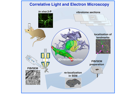
Label-free 3D-CLEM using endogenous tissue landmarks
We demonstrate feasibility of the workflow by combining in vivo 2-photon microscopy and focused ion beam scanning electron microscopy (FIB/SEM) to dissect the role of astrocytic coverage in the persistence of dendritic spines.
Emerging 3D correlative light and electron microscopy (CLEM) approaches enable studying neuronal structure-function relations at unprecedented depth and precision. However, established protocols for the correlation of light and electron micrographs rely ... Read more
Manja Luckner,Steffen Burgold, Severin Filser, Maximilian Scheungrab, Yilmaz Niyaz, Eric Hummel, Gerhard Wanner, Jochen Herms
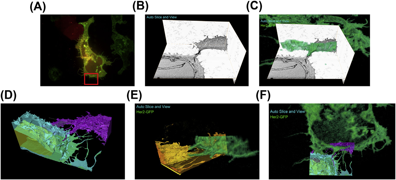
A fully integrated, three-dimensional fluorescence to electron microscopy correlative workflow
While fluorescence microscopy provides tools for highly specific labeling and sensitive detection, its resolution limit and lack of general contrast has hindered studies of cellular structure and protein localization. Recent advances in correlative light and electron microscopy (CLEM), including the fully integrated CLEM workflow instrument, the Thermo Scientific CorrSight with MAPS, have allowed for a more reliable, reproducible, and quicker approach to correlate three-dimensional time-lapse... Read more
Claudia S. Lopez, Cedric Bouchet-Marquis, Christopher P. Arthur, Jessica L. Riesterer, Gregor Heiss, Guillaume Thibault, Lee Pullan, Sunjong Kwon, Joe W. Gray
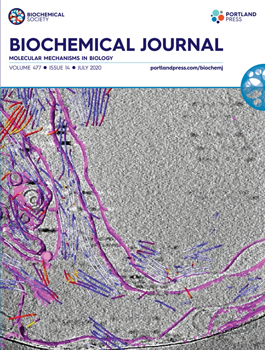
Synergistic role of nucleotides and lipids for the self-assembly of Shs1 septin oligomers
Amira capacities for membranes and filaments segmentation in cryo-TEM images are featured on the front cover of Biochemical Journal, July 2020.
Budding yeast septins are essential for cell division and polarity. (…) [The authors] have dissected, here, for the first time, the behavior of the Shs1 protomer bound to membranes at nanometer resolution, in complex with the other septins. Using electron microscopy, [the authors] have shown that on membranes, Shs1 protomers self-assembl... Read more
Cyntia Taveneau, Rémi Blanc, Gerard Pehau-Arnaudet, Aurélie Cicco, Aurélie Bertin
Ovipositor of the braconid wasp Habrobracon hebetor: structural and functional aspects
The Braconidae are a megadiverse and ecologically highly important group of insects. The vast majority of braconid wasps are parasitoids of other insects, usually attacking the egg or larval stages of their hosts. The ovipositor plays a crucial role in the assessment of the potential host and precise egg laying. We used lightand electron-microscopic techniques to investigate all inherent cuticular elements of the ovipositor (the female 9th abdominal tergum, two pairs of valvifers, and three p... Read more
Michael Csader, Karin Mayer, Oliver Betz, Stefan Fischer, Benjamin Eggs

Protocols for Generating Surfaces and Measuring 3D Organelle Morphology Using Amira
High-resolution 3D images of organelles are of paramount importance in cellular biology. Although light microscopy and transmission electron microscopy (TEM) have provided the standard for imaging cellular structures, they cannot provide 3D images.
However, recent technological advances such as serial block-face scanning electron microscopy (SBF-SEM) and focused ion beam scanning electron microscopy (FIB-SEM) provide the tools to create 3D images for the ultrastructural analysis of org... Read more
Edgar Garza-Lopez, Zer Vue, Prasanna Katti, Kit Neikirk, Michelle Biete, Jacob Lam, Heather K. Beasley, Andrea G. Marshall, Taylor A. Rodman, Trace A. Christensen, Jeffrey L. Salisbury, Larry Vang, Margaret Mungai, Salma Ash Shareef, Sandra A. Murray, Jianqiang Shao, Jennifer Streeter, Brian Glancy, Renata O. Pereira1, E. Dale Abel, and Antentor Hinton, Jr.
The molecular basis for sarcomere organization in vertebrate skeletal muscle
Sarcomeres are force-generating and load-bearing devices of muscles. A precise molecular picture of how sarcomeres are built underpins understanding their role in health and disease. Here, we determine the molecular architecture of native vertebrate skeletal sarcomeres by electron cryo-tomography.
Our reconstruction reveals molecular details of the three-dimensional organization and interaction of actin and myosin in the A-band, I-band, and Z-disc and demonstrates that α-actinin cros... Read more
Zhexin Wang, Michael Grange, Thorsten Wagner, Ay Lin Kho, Mathias Gautel, Stefan Raunser
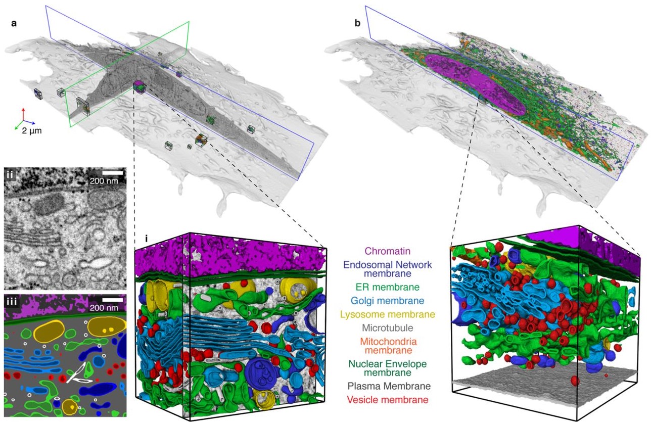
Automatic whole cell organelle segmentation in volumetric electron microscopy
Cells contain hundreds of different organelle and macromolecular assemblies intricately organized relative to each other to meet any cellular demands. Obtaining a complete understanding of their organization is challenging and requires nanometer-level, three-dimensional reconstruction of whole cells. Even then, the immense size of datasets and large number of structures to be characterized requires generalizable, automatic methods.
To meet this challenge, we developed an analy... Read more
Larissa Heinrich, Davis Bennett, David Ackerman, Woohyun Park, John Bogovic, View ORCID ProfileNils Eckstein, Alyson Petruncio, Jody Clements, C. Shan Xu, Jan Funke, Wyatt Korff, Harald F. Hess, Jennifer Lippincott-Schwartz, Stephan Saalfeld, Aubrey V. Weigel, COSEM Project Team
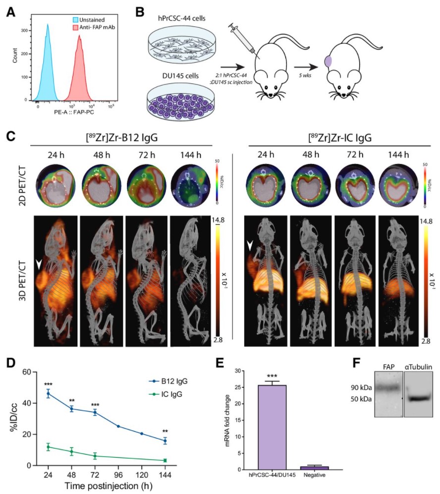
Malignant cells are surrounded by a complex and supportive tumor microenvironment that consists of immune cells, extracellular matrix, vasculature, and fibroblasts. Cancer-associated fibroblasts (CAF) are the major cell type in the reactive stroma and are known to promote tumorigenesis and metastasis. Fibroblast activation protein alpha (FAP) is a transmembrane serine protease expressed by CAFs in the microenvironment of epithelial tumors.
Meta-analysis of FAP expression and clinical ... Read more
Hallie M. Hintz, Joseph P. Gallant, Donald J. Vander Griend, Ilsa M. Coleman, Peter S. Nelson and Aaron M. LeBeau
Osteocytes are the most frequent bone cells connected with each other through cell processes within tiny tubular-shaped canaliculi. The so-called osteocyte lacunar-canalicular network (LCN) plays a crucial role in bone remodeling and mineral homeostasis. Given the critical nature of these functions, it is herein hypothesized that the LCN must be structurally “overengineered” to provide network resilience.
This hypothesis is tested by characterizing canalicular networks in human bon... Read more
Emely Bortel, Liam M Grover, Neil Eisenstein, Christian Seim, Heikki Suhonen, Alexandra Pacureanu, Peter Westenberger, Kay Raum, Max Langer, Francoise Peyrin, Owen Addison, Bernhard Hesse
Precise methods for quantifying drug accumulation in brain tissue are currently very limited, challenging the development of new therapeutics for brain disorders. Transcardial perfusion is instrumental for removing the intravascular fraction of an injected compound, thereby allowing for ex vivo assessment of extravasation into the brain. However, pathological remodeling of tissue microenvironment can affect the efficiency of transcardial perfusion, which has been largely overlooked.
We... Read more
Serhii Kostrikov, Kasper B. Johnsen, Thomas H. Braunstein, Johann M. Gudbergsson, Frederikke P. Fliedner, Elisabeth A. A. Obara, Petra Hamerlik, Anders E. Hansen, Andreas Kjaer, Casper Hempel & Thomas L. Andresen
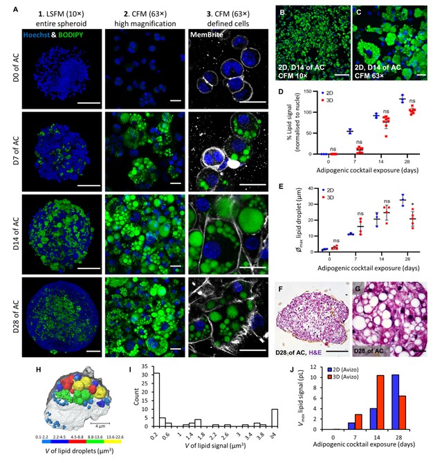
Adipose models have been applied to mechanistic studies of metabolic diseases (such as diabetes) and the subsequent discovery of new therapeutics. However, typical models are either insufficiently complex (2D cell cultures) or expensive and labor intensive (mice/in vivo). To bridge the gap between these models and in order to better inform pre-clinical studies we have developed a drug-responsive 3D model of white adipose tissue (WAT).
Here, spheroids (680 ± 60 μm) comp... Read more
Alexander D Graham, Rajesh Pandey, Viktoriya S Tsancheva, Alessia Candeo, Stanley W Botchway, Alasdair J Allan, Lydia Teboul4, Kamel Madi, Tahkur S Babra, Louisa A K Zolkiewski, Xuan Xue, Liz Bentley, Joan Gannon, Sam N Olof and Roger D Cox